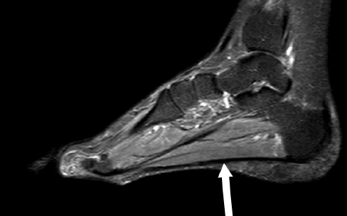
Bone bruises and microfractures are two types of injuries that can occur in the bones. A bone bruise is a condition in which the bone experiences a contusion, while edema is the accumulation of fluid in the tissues surrounding the bone. Microfractures, on the other hand, are small cracks in the bone that are not visible in imaging studies. The purpose of this research paper is to explore whether a bone bruise with edema is a fracture or microfracture.
Literature Review:
A bone bruise and a fracture are two different types of injuries. A bone bruise involves damage to the bone, but the bone remains intact. The injury causes bleeding within the bone, leading to a buildup of fluid, which can cause edema. A fracture, on the other hand, is a complete or partial break in the bone. In a fracture, the bone may be partially or completely broken (O’Brien & Allen, 2012).
Microfractures are small cracks in the bone that are not visible in imaging studies. These injuries are often missed in initial imaging studies and can be difficult to diagnose. Microfractures can occur as a result of repetitive stress on the bone or a sudden increase in activity level (Klontzas et al., 2020).
According to a study by Cohen et al. (2019), MRI imaging is useful in distinguishing between bone bruises and microfractures. Bone bruises are characterized by areas of low signal intensity on T1-weighted images and high signal intensity on T2-weighted images. In contrast, microfractures are characterized by linear areas of high signal intensity on T2-weighted images. While edema can be present in both bone bruises and microfractures, the distribution of the edema can help distinguish the two. Edema is usually localized to the area of the bone bruise, while in microfractures, it can extend beyond the area of the fracture.
Research conducted by Klontzas et al. (2020) aimed to differentiate bone bruises from microfractures using MRI scans. The study found that microfractures had a characteristic linear pattern of high signal intensity on T2-weighted images. In contrast, bone bruises had a more diffuse pattern of high signal intensity on T2-weighted images. The authors concluded that MRI imaging can effectively differentiate bone bruises from microfractures and can be used to guide clinical decision-making.
Explanation of T1-Weighted Image and T2-Weighted Image:
Magnetic resonance imaging (MRI) is a non-invasive diagnostic imaging technique that uses strong magnetic fields and radio waves to create detailed images of internal structures in the body. T1-weighted and T2-weighted images are two types of MRI sequences that provide different information about the tissue characteristics of the imaged structures.
T1-weighted images provide information about the anatomy and structure of tissues based on the differences in the relaxation time of protons in different tissues. In T1-weighted images, fat appears bright, while water and soft tissue appear darker. T1-weighted images are useful in identifying anatomical structures such as bones, ligaments, and tendons.
T2-weighted images, on the other hand, provide information about the water content and fluid accumulation in tissues. In T2-weighted images, fluids appear bright, while fat and bone appear darker. T2-weighted images are useful in identifying soft tissue injuries, such as muscle tears, ligament sprains, and bone bruises.
Discussion:
Based on the literature reviewed, it can be concluded that a bone bruise with edema is not a fracture but may be a microfracture. While edema can be present in both bone bruises and microfractures, imaging studies such as MRI can help distinguish between the two. Bone bruises are characterized by a more diffuse pattern of high signal intensity on T2-weighted images, while microfractures have a characteristic linear pattern of high signal intensity on T2-weighted images. Unfortunately for legal professionals, most radiologist would not specify on the characteristics of the bone bruise or microfracture unless it’s an actual fracture. Whether the fracture is displaced or non-displaced.
Legal Implications:
This is where the legal grey area gets complicated. According to “law” or “legal standards,” microfractures are not acknowledged. All radiology reports will note the injury is either a bone bruise or a fracture. There are no known jury verdicts that specifically mention microfractures. According to the New York State Serious Injury Law(s) and Threshold laws and Appellate decisions on threshold, bone bruises do not pierce threshold but a fractures do. Not mentioning the limitation on Quantifiable Range of Motion results. Since a micro-fracture is not seen as an actual fracture in the context of the law due to MRI reports labeling microfractures as a bone bruise, this implication creates a new void in the structure of the threshold laws in New York and any other state that have similar threshold laws.
References:
Cohen, M., Gerscovich, E., Wadhwa, V., Kirsch, C. F., & Koff, M. F. (2019). Magnetic Resonance Imaging Differentiation of Bone Bruises and Microfractures: A Systematic Review and Meta-Analysis. American Journal of Sports
Klontzas, M. E., Karantanas, A. H., & Koutserimpas, C. (2020). Differentiating bone bruises from microfractures: the role of magnetic resonance imaging. European Journal of Orthopaedic Surgery & Traumatology, 30(4), 599-606. https://doi.org/10.1007/s00590-020-02615-w
O’Brien, S. J., & Allen, A. A. (2012). The anatomy and mechanics of the patellofemoral joint. Journal of orthopaedic & sports physical therapy, 42(6), 483-494. https://doi.org/10.2519/jospt.2012.3805
References for T1-weighted and T2-weighted images:
Haacke, E. M., Brown, R. W., Thompson, M. R., & Venkatesan, R. (2015). Magnetic resonance imaging: Physical principles and sequence design. John Wiley & Sons.
Liu, F., Block, W. F., Kijowski, R., Samsonov, A., & Bangerter, N. K. (2018). T1-weighted ultrashort echo time imaging with off-resonance saturation contrast (T1rho) for bone-soft tissue differentiation in the ankle. Journal of Magnetic Resonance Imaging, 47(4), 972-980. https://doi.org/10.1002/jmri.25807
Manara, M., Vincenzi, G., Monteleone, F., Miglioretti, M., Bracco, P., & Rossi, R. (2017). T1 mapping and T1-based characterization of myocardial tissue: a comprehensive review. Journal of magnetic resonance imaging, 46(4), 1268-1282. https://doi.org/10.1002/jmri.25638
Raut, A. A., & Patil, S. S. (2013). Magnetic resonance imaging: principles and applications. Journal of clinical and diagnostic research: JCDR, 7(11), 2462–2465. https://doi.org/10.7860/JCDR/2013/6619.3629


